
X-rays are one of the most widely used medical imaging tools in hospitals and clinics. They help healthcare providers diagnose and monitor a variety of conditions. While X-rays are most associated with broken bones or dental exams, they also provide valuable imaging for detecting infections, tumors, and internal injuries. But how do X-rays operate, and what kind of information can they reveal about the human body? This article looks at the science behind X-rays and how medical professionals use and interpret these images.
What Is an X-Ray?
An X-ray is a type of electromagnetic radiation that may pass through the body and create images of its inside components. Unlike visible light, which is absorbed or reflected by objects, X-rays have higher energy levels and can penetrate tissues at varying rates. Dense materials, such as bones, absorb more X-rays and appear white in an X-ray image, whereas softer tissues absorb less and show in shades of grey.
First discovered in 1895 by German physicist Wilhelm Conrad Roentgen, he called it X-radiation, to represent its unknown nature. This mysterious radiation had the ability to pass through many materials that absorb visible light. These days, healthcare providers use X-ray imaging to examine bones, organs, and soft tissues, allowing non-invasive glimpses inside the body making it a crucial diagnostic tool.
How Do X-Rays Work?
X-ray machines produce a controlled beam of radiation that travels through the body and is picked up by a detector or film on the other side. The resulting image relies on the amount of radiation absorbed by different tissues:
- Bones absorb the most radiation and appear white.
- Muscles and soft tissues absorb less and appear in shades of gray.
- Air-filled spaces, such as the lungs, absorb the least and appear black.
Modern digital X-ray systems use detectors instead of traditional film, allowing for quicker image processing and digital storage. Additionally, advancements in imaging technology have significantly reduced the amount of radiation exposure patients receive during an X-ray examination.
Different Types of X-Rays
X-ray imaging technology has evolved, leading to various types of X-rays designed for specific medical applications. Some of the most common include:
- Radiography: The standard form of X-ray imaging, used to detect fractures, infections, and lung conditions like pneumonia.
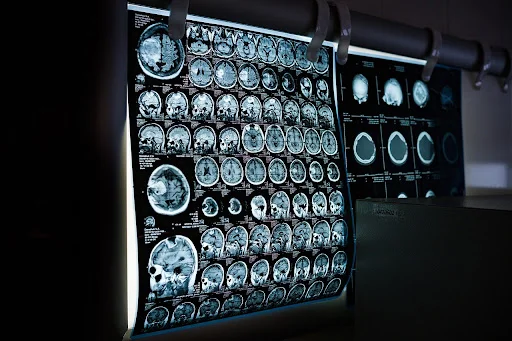
- Computed Tomography (CT) Scans: A series of X-ray images taken from different angles and compiled into a 3D image, often used for diagnosing tumors, internal bleeding, and organ damage.
- Fluoroscopy: A real-time X-ray that captures motion, often used in guiding procedures like catheter insertions or digestive tract examinations.
- Mammography: A specialized X-ray used for detecting breast cancer by capturing detailed images of breast tissue.
- Dental X-Rays: Dentists use X-rays to assess tooth decay, infections, and bone structure in the jaw.
Each type of X-ray has specific applications and benefits that allow healthcare providers to choose the most appropriate imaging method for their patients.
What Can an X-Ray Show?
X-rays provide detailed images of various parts of the body, helping doctors to detect and diagnose numerous conditions, including:
- Bone fractures and joint dislocations: X-rays are widely used in emergency rooms to assess breaks, fractures, and dislocated joints
- Infections: Pneumonia, tuberculosis, and other infections can be detected through chest X-rays
- Arthritis: X-ray image can reveal joint damage and the progression of conditions like osteoarthritis
- Tumors and cancer: CT scans and mammograms help detect abnormal growths such as lipomas and potential cancerous masses
- Lung and heart conditions: Chest X-rays can identify lung diseases, fluid buildup, or heart enlargement
- Foreign objects: X-rays are useful in locating swallowed or embedded foreign objects in the body
By revealing abnormalities that may not be visible externally, X-rays enable doctors to diagnose patients sooner and begin treatment for many medical conditions a lot earlier.
Who Can Read X-Rays?
Interpreting X-ray images requires specialized training, and only qualified healthcare professionals are allowed to read these images. Radiologists, who are medical doctors specializing in imaging, are the primary experts in reading and diagnosing conditions from X-rays. Trained healthcare providers, such as nurse practitioners and physician assistants, can also interpret X-ray results in clinical settings.
Nurses who have completed an online MSN FNP program receive training in ordering and interpreting diagnostic tests, including X-rays. These nurses play an important role in patient care by identifying medical conditions and determining appropriate treatment plans based on imaging results.
Understanding X-Rays Saves Lives
X-rays are a powerful and essential tool in modern medicine, allowing healthcare providers to diagnose and treat a wide range of conditions. From detecting broken bones to identifying life-threatening diseases, X-ray technology continues to play a vital role in healthcare and medical diagnostics. As imaging techniques advance, X-rays are becoming safer, more detailed, and more accessible, improving patient care across various medical fields. Whether analyzed by radiologists or qualified nurses, X-rays are a valuable resource for all medical professionals.